Microscopic Enhanced RCT
Up to 25x magnification
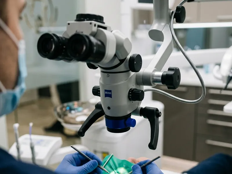
Root canals are incredibly small — often narrower than a human hair — and hidden deep inside the tooth. Microscopic Enhanced RCT uses a high-powered dental operating microscope to magnify the treatment area up to 25 times and illuminate it with intense fibre-optic light. The endodontist can look directly inside the canal system, locating hidden structures, micro-cracks and complex anatomy invisible to the naked eye.
Micro-surgical endodontics by specialists
Led by Dr Rashmi P. Patil, Shwetha Dentistry features an advanced dental operating microscope. We specialise in complex micro-endodontics, including retrieving broken dental files, treating calcified roots and resolving failed root canals (retreatments).
Surgical microscope (25x)
Visualises hidden root canals, extra roots and micro-fractures.
Micro-ultrasonic instruments
Cleans canals with microscopic precision without weakening the tooth.
Failed-RCT retreatments
Saves teeth previously declared untreatable by locating missed canals.
Who is it for?
- Teeth with highly complex, curved or split root canal systems
- Patients needing retreatment of a previously failed root canal
- Teeth suspected of having micro-fractures or extra hidden canals
- Cases where a broken dental instrument needs to be retrieved
What to expect
The procedure is performed with a large surgical microscope positioned directly over your mouth. Our endodontist looks through the lenses to operate. The experience is highly comfortable, and the magnification ensures the utmost precision — often reducing the amount of drilling needed.
Performed at Shwetha Dentistry by our specialist endodontic team.
The Procedure, Step by Step
Exactly what happens during your microscopic enhanced rct — no surprises.
Microscope Alignment & Access
Microscope Alignment & Access
Positioning the surgical microscope and preparing a micro-access cavity.
The surgical microscope is positioned and adjusted over the patient. The endodontist prepares a micro-access cavity, keeping it as small as possible to conserve the tooth’s natural crown.
The microscope provides bright illumination and clear visibility from the start.
Orifice Location
Orifice Location
Locating all root canal openings under high magnification.
Under 10x to 25x magnification, the endodontist examines the pulp chamber floor to locate all canal openings — including tiny extra canals (like the MB2 canal in upper molars) that are often missed.
Locating every canal is critical to preventing root canal failure.
Micro-Cleaning
Micro-Cleaning
Cleaning the canals precisely using micro-instruments.
The canals are cleaned and shaped using fine micro-files and micro-ultrasonic tips. The magnification lets the endodontist remove only the infected tissue while conserving healthy dentine.
Precise cleaning maintains the structural strength of the root.
Micro-Crack Inspection
Micro-Crack Inspection
Inspecting the internal walls for micro-fractures or cracks.
The endodontist inspects the internal canal walls under high magnification to check for micro-cracks or fractures. If a crack is found, it helps determine whether the tooth can be saved.
This diagnostic capability prevents unnecessary treatment of cracked teeth.
Precision Micro-Obturation
Precision Micro-Obturation
Filling and sealing the canals under direct vision.
The clean canals are filled with a bioceramic sealer and gutta-percha. The endodontist visually verifies the adaptation and quality of the seal under the microscope before placing a temporary filling.
Direct visual confirmation ensures a complete, leak-proof seal.
Treatment Cost
₹7,500 – ₹9,500
1–2 visits (60–80 minutes per visit)
Indicative range — please consult the dentist; the exact price varies from case to case.
Aftercare
- Avoid chewing on the treated side until the final crown is placed.
- Maintain standard oral hygiene with brushing and flossing.
- Temporary soreness is normal; manage it with standard pain relievers.
- Follow up as scheduled to place the permanent crown.
Frequently Asked Questions
It is highly recommended for complex cases (like molars with 3 or 4 roots), calcified canals, retreatments of failed root canals, or when a broken instrument needs to be located.
Yes. At Shwetha Dentistry, microscopic treatments are performed by our specialist endodontist, Dr Rashmi P. Patil, who is trained in micro-surgical endodontics.
Because of the extreme detail and care involved, it can take slightly longer (around 60 to 90 minutes) and sometimes requires two sessions to ensure absolute cleanliness.
Dental loupes provide 2.5x to 4.5x magnification, while surgical microscopes provide up to 25x with coaxial fibre-optic lighting — revealing details loupes miss.
Yes. Under high magnification and illumination we can see the broken fragment and use specialised micro-ultrasonic tips to loosen and retrieve it safely.
Without magnification, finding hidden canals or resolving complications is difficult — microscopes are essential for these challenging cases.
Yes. Studies show microscopic RCT has a success rate of over 98% because it minimises the risk of missed canals, the main cause of failures.
No. The microscope remains well above your face and never touches you. Most patients find it relaxing because the doctor works quietly and efficiently.
It ranges from ₹7,500 to ₹9,500 depending on complexity — excellent value given the specialist skill and equipment involved. Please consult the dentist for an exact quote.
Yes, although it is most beneficial for multi-rooted molars, premolars and teeth undergoing retreatment.